
Patient-derived models, made in MOS
MOS start from the lab's own samples: cell lines, PDX, PDO, and biopsies, fresh or frozen. Using a single-cell suspension as input, MOSgen makes up to 280,000 MOS per run.
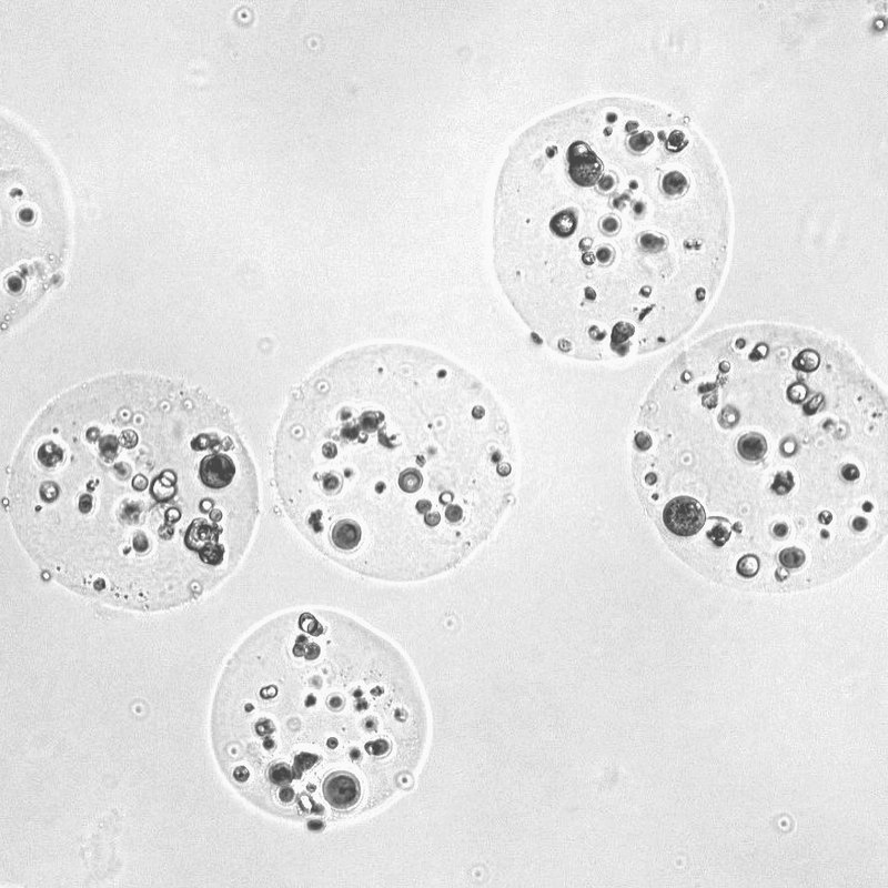
MOS in a well.
Tissues used in MOS
MOS have been made from cancer and normal tissue across nine organs, from fresh or frozen samples.
Lung
Non-small cell lung cancer (NSCLC) and normal lung.
About 50 PDOs and A549 cells established in MOS.
Breast
Breast cancer and normal breast.
About 40 PDOs and MCF7 cells established in MOS.
Colon
Colorectal cancer and normal colon.
About 35 PDOs and Caco2 cells established in MOS.
Bone marrow
Multiple myeloma and normal bone marrow.
MM1S and H929 cells established in MOS.
Skin
Melanoma and normal skin.
A375 cells established in MOS.
Liver
Normal liver, including primary human hepatocytes.
Pancreas
Pancreatic cancer (PDAC).
5 PDOs established in MOS.
Ovary
Ovarian cancer.
Prostate
Prostate cancer.
Source: Uniform Bio, Research with MOS.
What goes into a MOS
Each MOS is a miniaturized droplet of matrix with a selectable number of starting cells, set by a simple dilution. Cells proliferate and organize inside every MOS.
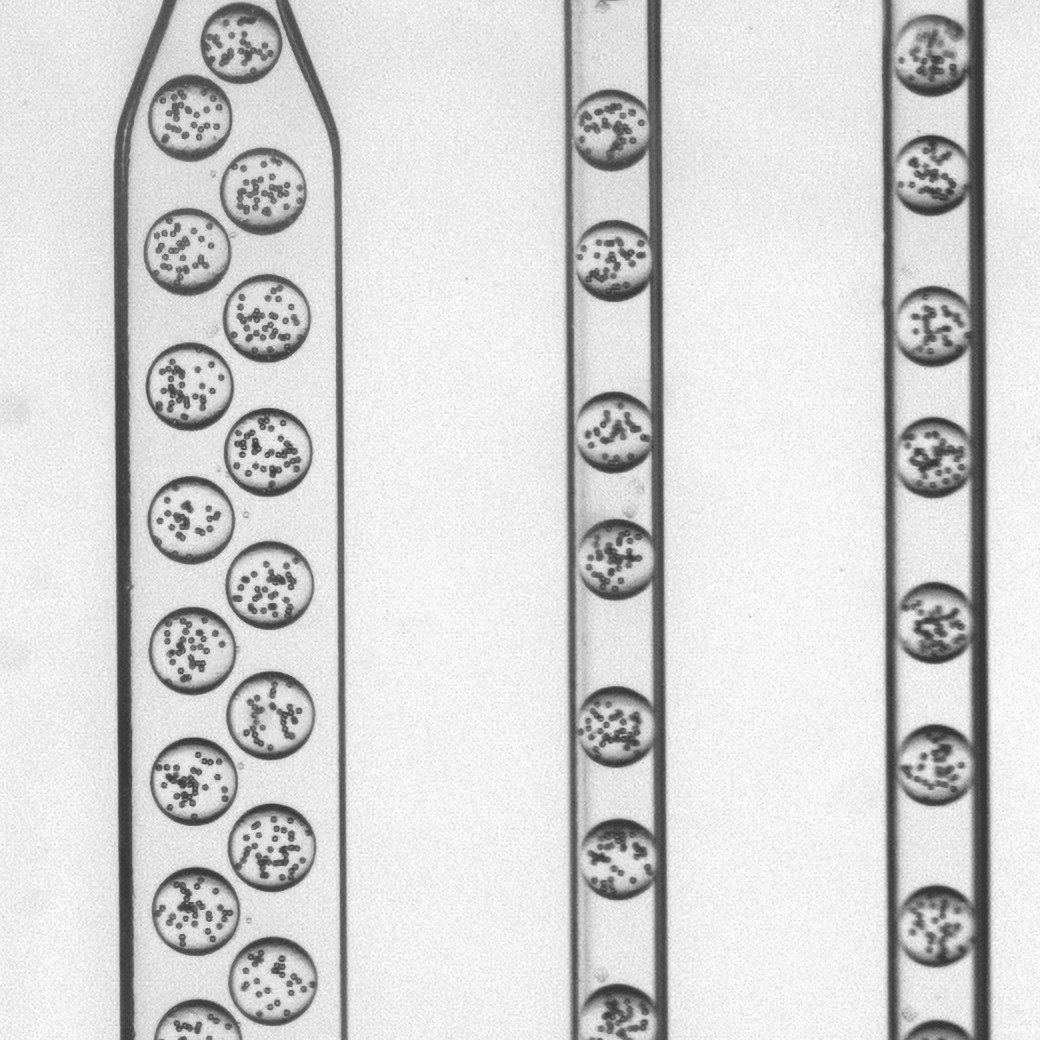
In the chip
Cells in matrix form droplets, four lanes at a time.
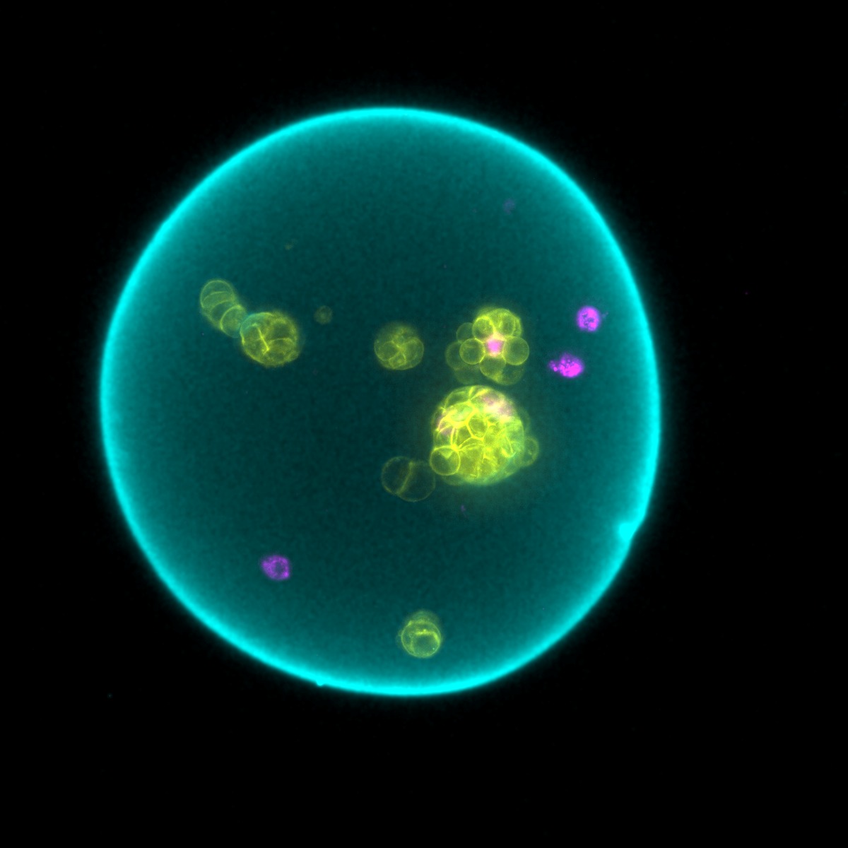
One MOS
A single MOS with organoids growing inside, imaged by fluorescence.
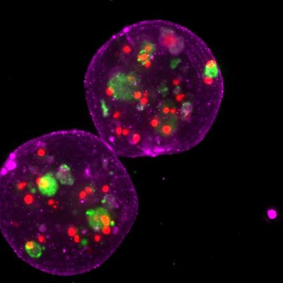
Imaged whole
MOS (magenta) containing live patient organoids.
Why MOS instead of hand-plated domes
The matrix in one hand-plated dome makes many MOS, and each MOS is its own data point.
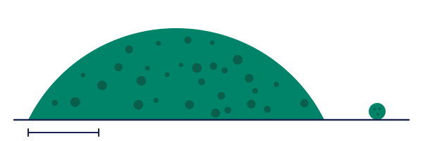
Drawn to scale: a 10 µL dome beside one MOS at the standard droplet size. The bar is 1 mm.
Sources: Gobits R, et al. JCO Precision Oncology, 2026 (reproducibility). Ding S, et al. Cell Stem Cell, 2022 (immune cell access). Schoonbeek MC, et al. EJC Paediatric Oncology, 2026 (sample per data point). Other rows: Uniform Bio data.
Bring your samples
Tell us your sample types and the assays you run, and we will plan an evaluation of MOSgen with you.